Allograft bio-implants
Overview
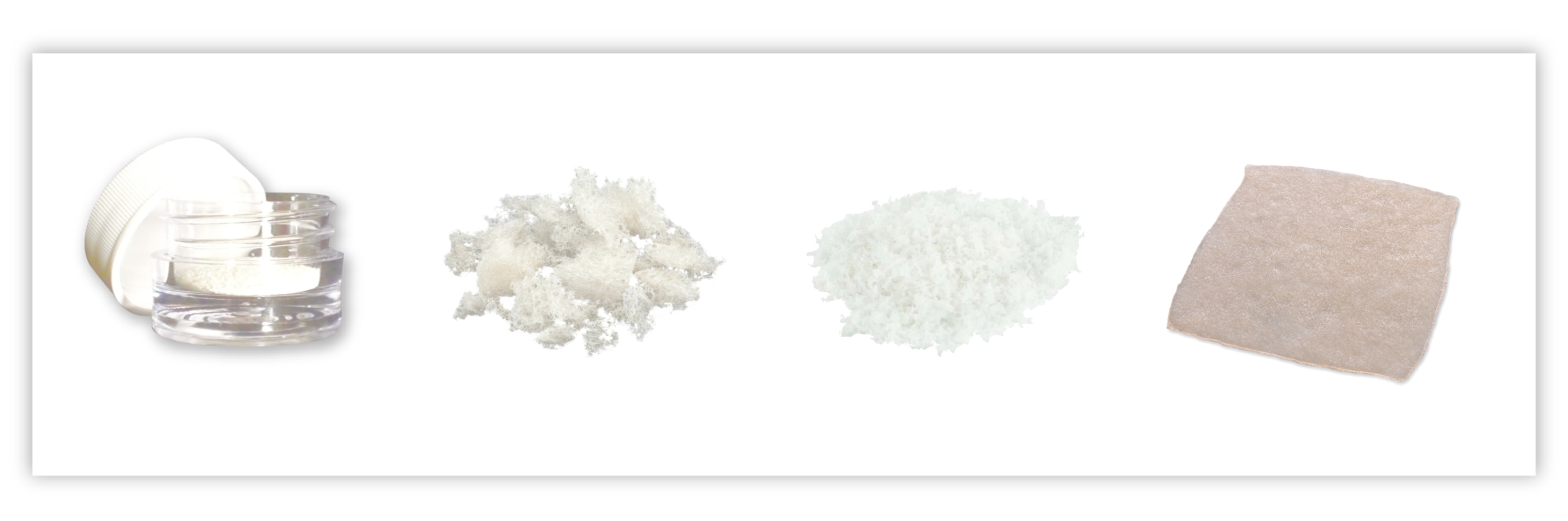
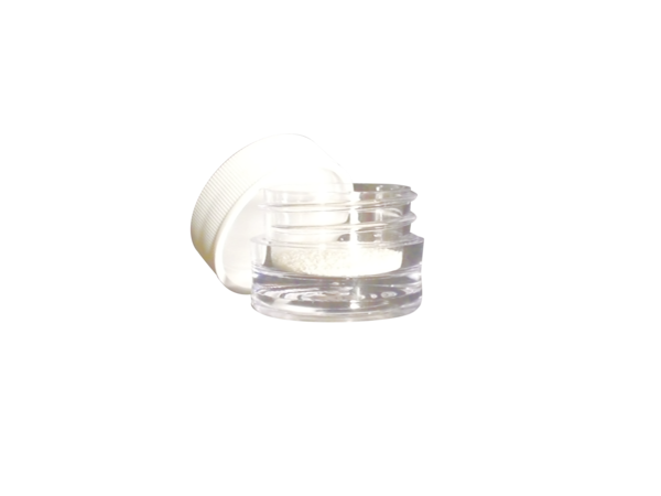
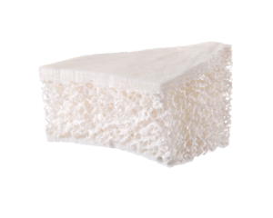
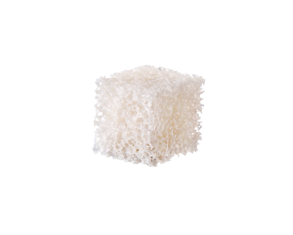
Our allograft bio-implants come in a variety of grafts and sizes to fit your Craniomaxillofacial needs. They have a SAL of 10-6, resulting in a potential infection rate of 1 in 1,000,000.1 The implants can be stored at room temperature, unfrozen, and ready when the surgeon needs it.
Features and benefits
Safe
SAL of 10-6, resulting in a potential infection rate of 1 in 1,000,000.1
Ready to use
Can be stored at room temperature, unfrozen, and ready when the surgeon needs it.
Comprehensive offering
Variety of grafts and sizes to choose from to fit your Craniomaxillofacial needs.
Clinical evidence
BACKGROUND
Demineralized freeze-dried bone allograft (DFDBA) is widely used in periodontal therapy as a scaffold for new bone formation in periodontal defects. It is demineralized, theoretically, to expose osteoinductive or osteoconductive bone matrix proteins that should facilitate osteogenesis. The degree of DFDBA demineralization varies between tissue banks and may affect clinical regeneration. A 2% residual calcium level in DFDBA has been shown to result in the highest alkaline phosphatase activity levels in cultured human periosteal cells and is optimally osteoinductive or osteoconductive for new bone formation. The purpose of this study was to evaluate the effect of 4 different residual calcium levels in commercially available DFDBA samples on porcine osteoclast activity as measured by resorption on calcium phosphate-coated disks.
METHODS
Bone marrow was harvested from the femurs of 3-week-old farm pigs and cultured for 3 weeks. Hematopoietic stem cells were allowed to differentiate into mature active polykaryons displaying genuine osteoclast characteristics. The osteoclast cells displayed a dense actin band inside the margins of the cytoplasm under light microscopy. Culture media was decanted and collagenase added to free the attached cells. Equal cell samples were pipetted onto calcium phosphate-coated disks in 24-well plates. DFDBA samples with 1.44%, 2.41%, and 5.29% residual calcium; FDBA (30% residual calcium); and control cultures without allograft samples were prepared and all samples incubated for 1 week. Cells were fixed and stained for tartrate-resistant acid phosphatase (TRAP), Oregon Green 488-phalloidin, a stain for cytoskeletal proteins, and counterstained with propidium iodide. Specimens were examined by light and fluorescence microscopy using epi-illumination. Calcium phosphate disks were then rinsed in 5% sodium hypochlorite to remove adherent osteoclasts, and substrate surface changes were measured by white light interferometry and image analysis.
RESULTS
A higher yield of TRAP-positive cells was produced without DFDBA; however, resorptive activity appears to be significantly increased in the presence of 2.41% residual calcium as compared to all other experimental groups (P<0.0065).
CONCLUSION
In this in vitro model, porcine osteoclasts show significantly more resorptive activity as measured on calcium phosphate-coated disks in the presence of 2.41% residual calcium in DFDBA than in other DFDBA residual calcium levels.
The relationships between residual calcium levels and particle size of ground demineralized bone matrix and its osteoinductive potential were investigated using in vitro and in vivo assays. The effects of variable residual calcium levels, variable particle sizes, and donor age and gender were studied using a tissue culture-based bioassay (in vitro) as well as an athymic mouse (in vivo) bioassay. The osteoinductive potential of the bone-derived biomaterial was assessed by measuring the degree of new bone formation (change in percent calcium content after 4 weeks of implantation) in the in vivo assay and levels of alkaline phosphatase activity associated with cultures of human periosteal cells (HPO cells) in the in vitro assay, respectively. Slightly demineralized bone matrix and overly demineralized bone matrix possessed a degree of osteoinductive potential whereas bone demineralized to levels of approximately 2% residual calcium provided for maximum osteoinductive potential in both assay systems. The osteoinductive potential of ground demineralized bone varied relative to the particle size such that DBM particles ranging from 500 to 710 microns provided for the highest level of calcium deposition (increase of 8.1 weight percent calcium) after 4 weeks of implantation in muscle pouches of an athymic mouse, whereas explanted particles less than 250 microns showed the lowest level of calcium deposition (increase of only 2.8 weight percent calcium). In the donor age and gender study, DBM from different donors were divided into 5 age groups for both female and male donor derived bone: less than 20, 21 to 30, 31 to 40, 41 to 50, and 51 to 60 year old age groups. This study indicated that DBM from female donors in the 31 to 40 years old age group and male donors in the 41 to 50 year age group possess the highest osteoinductive potential, whereas DBM derived from donor bone from both female and male donors in the 51 to 60 year age group presented the lowest osteoinductive potential. DBM derived from male and female donors did not in general show significant differences in osteoinductive potential.
Demineralized freeze-dried bone allograft (DFDBA), a widely used graft material in periodontal regenerative procedures, is processed with hydrochloric acid in the attempt to expose proteins located within the bone matrixes that are capable of inducing new bone formation. However, the degree of DFDBA demineralization varies between tissue banks, which may have an effect on clinical regeneration. This study uses the critical-sized defect (CSD) model to evaluate the wound-healing response to the residual calcium of donor bone. If the percentage of residual calcium in a graft were demonstrated to significantly enhance wound healing, then periodontal patients may benefit from further standardization of human-allograft processing. Sixty adult, male, Harlan Sprague-Dawley rats (Rattus norvegicus) were randomly and equally divided into 4 test groups (ie, DFDBA at 1%, 2%, and 3% to 6% residual calcium levels and FDBA at 23% residual calcium) and a control group (no allograft). An 8-mm-diameter craniotomy was made in the rat calvarium, and polytetrafluoroethylene membranes with pore sizes of 0.50 microm were placed intracranially and ectocranially. Treatment materials were carefully placed into the CSD with a new sterilized dental amalgam carrier. Tetracycline hydrochloride was injected intraperitoneally for labeling new bone growth, and animals were euthanized 12 weeks postsurgery. As a result, histomorphometric bone fill at 12 weeks showed a statistically significant increase in the 2% DFDBA group as compared to all other groups. The authors conclude that a 2% residual calcium level in human DFDBA appears to significantly (P < or = .05) enhance osseous wound healing in the rat calvarium.
References:
1: “Ensuring the Safety of Allograft Tissue”, white paper on file with Stryker Craniomaxillofacial.
CMF-WC-42_Rev. None_18981